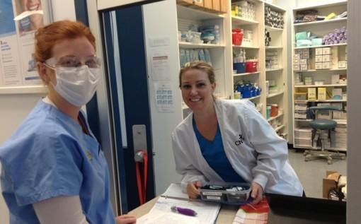
As ultrasonic technology developed, how did everyday work for sonographers change? As the medical field grows, ultrasonic technology has grown dramatically providing faster diagnosis, more accurate imaging, and better patient care. This page addresses the most current developments in ultrasonic technology—that which all ultrasound sonographers Canada should be familiar with. a will keep you current in an industry that is rapidly changing.
● Three- And Four-Dimensional Image Developments
Generation of 3D and 4D images is among the most important advances in ultrasonic technology. Three-dimensional pictures produced by 3D ultrasounds are clearer and more detailed than two-dimensional ultrasounds' produced flat images. This is considerably better with 4D ultrasounds, which provide time to the picture and generate real-time movie pictures.
● Little, Portable Ultrasonic Instruments
Portable and mobile ultrasonic equipment have transformed the sonographer's job. Apart from their ease of use, these little devices are remarkably durable. Ultrasound Sonographer Canada captures excellent photos and might be utilized at the patient's bedside or elsewhere.
● Integration Of Artificial Intelligence With Ultrasonic Imaging
Almost everywhere artificial intelligence (AI) is creating waves. There is not any difference in ultrasonic technology. In ultrasonic imaging, artificial intelligence integration refers to the use of machine learning techniques to improve picture quality, support result interpretation, and even automate certain testing process duties.
● Using Elastography: Gauging Tissue Stiffness
Elastography is a recently advanced and sophisticated ultrasonic method for tissue stiffness determination. This substantially helps to diagnose diseases like thyroid problems, breast cancer, and liver disease. By determining the tissue's stiffness, sonographers can give more all-encompassing information that would support early diagnosis and therapy planning.
● Improved Contrast Ultrasound
Another fascinating new invention about which sonographers should be aware is contrast-enhanced ultrasonic waves (CEUS). Under an ultrasonic examination, contrast drugs in CEUS aid to simplify blood flow and tissue vascularity visible. This method is very helpful in finding liver diseases, looking at renal masses, and keeping an eye on heart diseases.
●High Frequency Ultrasonic Waves Directed At Surface-Oriented Constructions
High frequency ultrasonic waves help to capture images of superficial tissues like the thyroid gland, breast tissue, and joint components. Higher frequencies provide more clarity, hence sonographers may find tiny problems not seen on lower-frequency ultrasounds.
Conclusion
Every sonographer should keep current as ultrasonic technology develops. Including portable devices, artificial intelligence, elastography, CEUS, high-frequency approaches to 3D and 4D imaging is changing medical imaging. Knowing how to use these contemporary technologies will enable a Canada ultrasonic sonographer to improve their work.
Looking for the best Ultrasound Sonographer Canada services? If yes, you can get it from a diagnostic medical sonography school in Ontario. Contact us at 613.726.CNIH(2644) or 1.866.726.CNIH(2644).